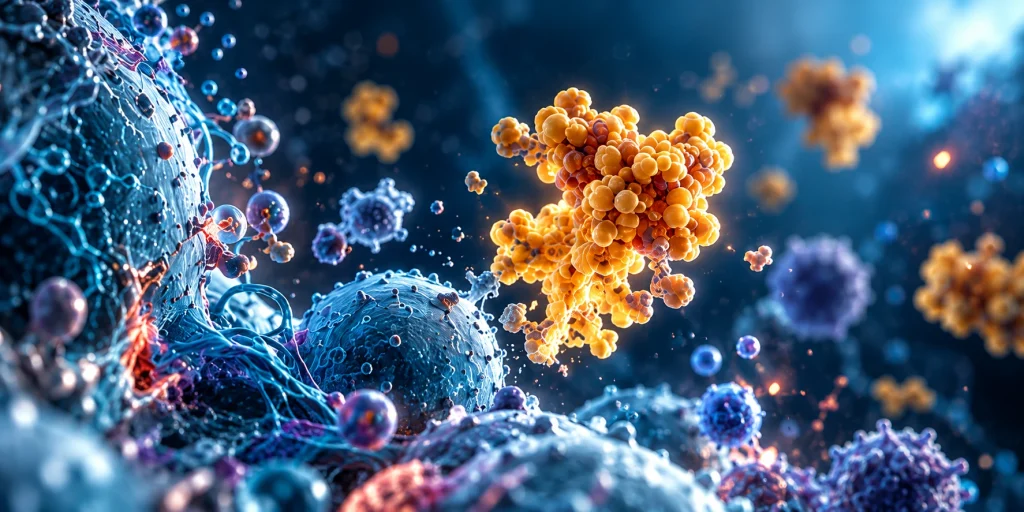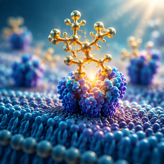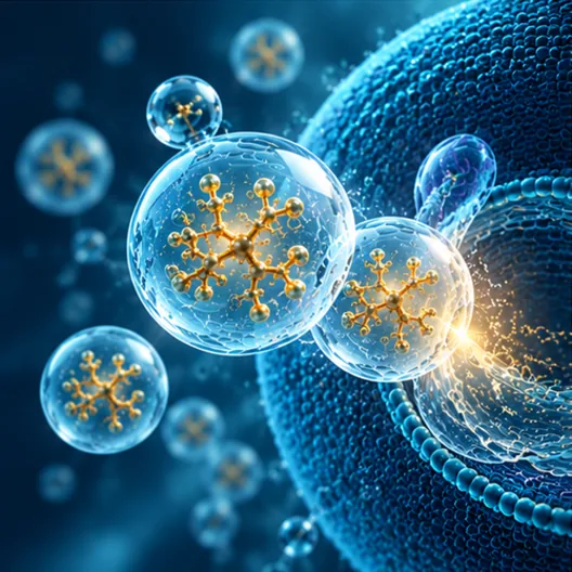

Over the past two decades, research has shown that elevated galectin-3 does more than mark disease—it helps drive it.
Heart Disease and Cardiac Fibrosis
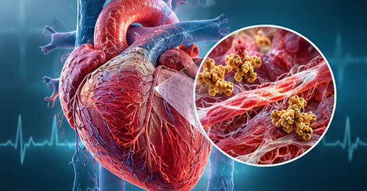
Activated macrophages in the heart secrete galectin-3, which tells fibroblasts to lay down excess collagen and scar tissue. That progressive fibrosis contributes to ventricular remodeling, reduced function, and heart failure. Circulating levels above 17.8 ng/mL are linked to higher risk of death or hospitalization in chronic heart failure, and galectin-3 is FDA-approved as a prognostic biomarker. It is also upregulated in unstable atherosclerotic plaques, where it amplifies inflammation and monocyte recruitment.
Inflammation and Immune Dysfunction
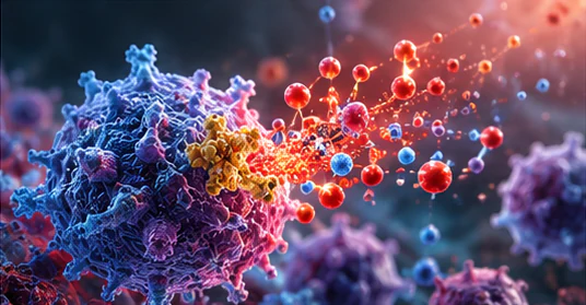
Galectin-3 acts as a master regulator of inflammation. It boosts pro-inflammatory cytokines (IL-1β, IL-6, TNF-α, IL-18) and can activate the NLRP3 inflammasome. When that pathway stays on, systemic inflammation and tissue injury follow. Reviews have tied it to cardiac, liver, kidney, and lung inflammation and fibrosis, as well as neuroinflammatory disease.
Organ Fibrosis
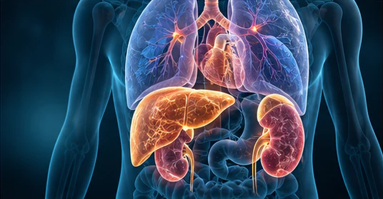
Its most damaging role may be fibrogenesis across organs:
- Liver fibrosis and NASH
- Kidney fibrosis and CKD progression
- Pulmonary fibrosis
- Cardiac fibrosis and heart failure
Knockout mice are protected from injury-driven fibrosis; animals with high galectin-3 develop severe scarring. Environmental toxins, including mold toxins, can raise galectin-3 and feed the same process.
Cancer Progression and Metastasis
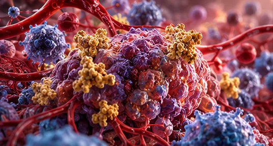
Many tumors overexpress galectin-3 and use it to evade immune surveillance, grow new blood vessels, migrate, and resist apoptosis and chemotherapy. Overexpression has been documented in breast, colon, thyroid, prostate, and ovarian cancers, and higher levels often track with more aggressive disease.
Neurodegeneration and Brain Aging
Activated microglia express galectin-3 and promote neuroinflammation. Elevated levels have been found in Alzheimer’s tissue and CSF, Huntington’s disease (correlating with severity), ALS plasma, and poor stroke outcomes. In Huntington’s models, galectin-3 accumulates at damaged lysosomes, blocks debris clearance, and fuels NF-κB and NLRP3 inflammation. Reducing it suppressed inflammation, lowered protein aggregation, and improved neuronal function and survival in mice.

Metabolic Dysfunction
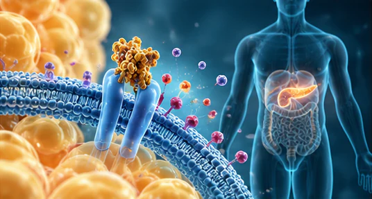
Extracellular galectin-3 can bind the insulin receptor and blunt insulin signaling. Knockout mice on a high-fat diet are protected from insulin resistance. High-fat diets also raise galectin-3 and activate TLR4/NLRP3 inflammatory pathways, linking chronic inflammation to metabolic disease.
Galectin-3 is both a useful biomarker and an active driver of fibrosis, inflammation, and tissue damage—which is why lowering or modulating it has become a research and clinical target.
A 2019 Nature Communications study found that galectin-3 builds up at damaged lysosomes in Huntington’s disease microglia. There, it blocks debris clearance and drives inflammation through NF-κB and the NLRP3 inflammasome. Knocking galectin-3 down reduced inflammation and protein aggregation, restored neuronal function, and improved survival in mouse models.